TL;DR: Your ear anatomy has three regions: the outer ear, middle ear, and inner ear. Sound crosses all three, and the inner ear makes nerve signals from vibration. Trouble anywhere along that chain changes what you hear.
Ear anatomy sounds like a classroom topic until your own hearing shifts. Every part of the ear holds one job, then hands sound along. Learning that chain makes hearing loss basics much less confusing. This guide walks the whole path, from the visible ear to the hearing nerve.
The Three Regions of Ear Anatomy at a Glance
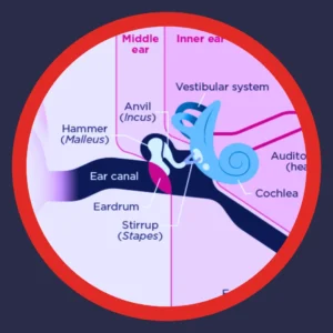
Hearing starts outside your head and finishes inside your brain. Three regions handle that trip, and each works in a different material. The table below sorts those regions into three neighborhoods.
|
Region |
Key structures |
Main job |
Works through |
|---|---|---|---|
|
Outer ear |
Pinna, ear canal, outer eardrum surface |
Gathers sound and funnels it inward |
Air |
|
Middle ear |
Eardrum, malleus, incus, stapes, eustachian tube |
Amplifies vibration and passes it along |
Air and bone |
|
Inner ear |
Cochlea, hair cells, semicircular canals, hearing nerve |
Turns motion into nerve signals |
Fluid |
Each handoff matters, because one weak link dulls everything that follows.
Sound changes form twice on this trip. Air pressure becomes physical movement in the middle ear. Movement then becomes fluid waves, and finally electricity. That double translation explains why hearing difficulty appears in many ways.
The Outer Ear: What You See and What You Do Not
Most people picture only the part they can pinch. The outer ear covers three pieces, and two hide inside your head. Together they collect sound and protect everything deeper.
The Pinna Shapes Sound Before You Hear It
That visible ridged funnel carries the name pinna, or auricle. Its folds are not decoration. They bounce and delay sound slightly, and your brain reads direction from that. Without that shaping, a footstep behind you would sound beside you.
The pinna earns its keep in a few specific ways:
- Collects sound waves across a wider area than a bare opening would
- Boosts speech frequencies slightly, right where conversation lives
- Adds direction cues your brain uses to place a sound
- Shields the canal opening from wind, water, and debris
Those four jobs run with no effort from you.
The Ear Canal Is a Tuned Tunnel
Your ear canal runs about an inch inward and bends slightly. The bend protects the eardrum from pokes and pressure. Skin in the outer third grows fine hairs and wax glands. That wax does real work.
Ear wax traps dust, repels water, and keeps canal skin supple. Problems start only when it packs tightly against the eardrum. Most questions about how to unclog your ear trace back to this tunnel. Dry canal skin causes itchy ears instead, and cotton swabs worsen it.
A few habits keep this part of your ear comfortable:
- Let wax migrate out on its own during chewing and talking
- Wipe only the outer bowl with a damp cloth
- Skip cotton swabs, hairpins, and ear candles entirely
- Dry your ears gently after swimming or a long shower
- Ask a provider about safe removal when hearing feels blocked
Small habits protect a small space. Your eardrum sits just beyond it.
The Middle Ear: A Bridge Built From Three Tiny Bones
Behind the eardrum sits an air-filled room the size of a pea. This pocket holds the smallest bones in the human body. Its whole purpose involves turning gentle air movement into real force.
The Eardrum Starts the Handoff
Your eardrum, or tympanic membrane, stretches thin and taut. Sound waves push it in and out thousands of times per second. The membrane copies those pressure changes almost perfectly. Even a pinhole dulls that copy and softens what you hear.
Malleus, Incus, and Stapes Do the Amplifying
Three linked bones cross that little room, and each has a nickname. Their chain multiplies force, because a wide eardrum pushes a small window. Ear anatomy solves a real physics problem right here.
|
Bone |
Common name |
Size and place |
Role in the chain |
|---|---|---|---|
|
Malleus |
Hammer |
Attaches to the eardrum |
Picks up eardrum movement |
|
Incus |
Anvil |
Sits in the middle |
Passes force to the stapes |
|
Stapes |
Stirrup |
Smallest bone you have |
Taps the inner ear window |
That three-bone lever adds the push that fluid would otherwise swallow.
Two tiny muscles also anchor to these bones. They tighten reflexively during sudden loud noise. Protection like that helps, though it reacts slowly for a gunshot.
The Eustachian Tube Keeps Pressure Even
A narrow tube links the middle ear to your throat. Swallowing and yawning open it briefly, which equalizes pressure across the eardrum. Airplane descents and head colds interrupt that balance. Hearing dulls until the tube opens again. Children have shorter, flatter tubes, so fluid lingers more easily.
Signs that this small tube needs attention include:
- Fullness that comes and goes with altitude or weather
- Crackling or popping when you swallow
- Hearing that muffles during and after a cold
- Discomfort that lingers days after a flight
Patterns like these point toward pressure, not permanent inner ear change.

The Inner Ear: Where Sound Becomes a Signal
Deep in the temporal bone sits the most delicate section. Fluid fills it, and two systems share the space. One handles hearing, and the other handles balance.
Inside the Cochlea
Picture a snail shell about the size of a lima bean. That spiral holds the cochlea, and fluid fills its coiled channels. When the stapes taps the oval window, waves ripple through that fluid. A flexible strip called the basilar membrane rides those waves.
Position along the strip decides pitch. Waves near the entrance carry high frequencies, and the far end takes low ones. Age-related change often hits high frequencies first. Birdsong and the letter S fade early.
Hair Cells Translate Motion Into Nerve Signals
Thousands of sensory cells line the basilar membrane. Each one wears a bundle of microscopic projections called stereocilia. Fluid motion bends those bundles, and the cell fires a chemical message. Federal research on hearing describes that bend as the moment sound becomes electricity.
Hair cells never grow back in humans. Three main forces damage them:
- Loud sound, especially repeated exposure over years
- Certain ototoxic medications that reach inner ear fluid
- Natural aging of the cells and their supporting structures
Protecting these cells beats repairing them, because repair is not possible.
Balance Shares the Same Address
Three looping semicircular canals sit beside the cochlea. Fluid inside shifts as your head turns, and sensors report that motion. Two small organs nearby track gravity and straight-line movement. Shared plumbing explains why dizziness and hearing changes often arrive together. Meniere’s disease works that way.
Balance clues worth mentioning at an appointment include:
- Spinning sensations that last minutes rather than seconds
- Unsteadiness that worsens in the dark
- Fullness in one ear alongside dizziness
Notes like these help a provider see the whole picture.
How Sound Travels Through Ear Anatomy, Step by Step
One conversation crosses every structure above in a fraction of a second. Walking the sequence once makes the whole map click. Follow a single word from your friend’s mouth to your understanding.
- Sound waves leave the speaker and reach your pinna.
- The pinna funnels those waves into your ear canal.
- Waves strike the eardrum, and it vibrates.
- Malleus, incus, and stapes carry that vibration across the middle ear.
- The stapes taps the oval window and starts fluid waves.
- Hair cells bend, then fire electrical signals.
- The hearing nerve delivers those signals to your brain for meaning.
Seven steps, and no conscious effort from you.
What Ear Anatomy Reveals About Hearing Loss
Location tells a provider a great deal. Where the trouble sits shapes both the experience and the options. Location also explains why two people describe hearing difficulty so differently.
|
Region affected |
Type of change |
What it often feels like |
Common contributors |
|---|---|---|---|
|
Outer or middle ear |
Conductive |
Volume drops, clarity holds up |
Wax blockage, fluid, eardrum injury |
|
Inner ear or nerve |
Sensorineural |
Words blur even at good volume |
Noise, aging, some medications |
|
Both regions |
Mixed |
Softer and less clear together |
Overlapping causes |
Sorting a change this way guides the next step.
Volume and clarity behave like separate dials. Conductive change turns the volume dial down, and many causes respond well. Inner ear change scrambles detail instead. Hearing aids then reshape sound for the cells that remain. Reading about conductive and sensorineural loss side by side sharpens that contrast. Degrees of hearing loss adds the measurement side.
Scale matters too. National hearing statistics put disabling hearing loss at 22% of adults 65 to 74. That share climbs to 55% after age 75. Numbers like those turn a private worry into an ordinary part of aging.
Matching Everyday Ear Sensations to the Right Region
Symptoms rarely announce their address. Still, several familiar sensations line up with specific regions. The table pairs common experiences with the structures most often involved.
|
What you notice |
Region usually involved |
Worth knowing |
|---|---|---|
|
Muffled hearing after a cold |
Middle ear and eustachian tube |
Pressure and fluid often clear with time |
|
A blocked, full feeling |
Outer ear canal |
Packed wax is a frequent cause |
|
Blood vessels near the middle ear |
A provider can help sort out the pattern |
|
|
Ringing after a concert |
Inner ear hair cells |
Repeated exposure adds up over years |
|
Dizziness with ear fullness |
Semicircular canals |
Balance and hearing share one space |
No pattern here replaces an exam, though each makes conversation easier.
Plain language about sensation helps more than guessing at a diagnosis. Describe when it started, which ear, and what helps. Details like those narrow the search quickly.

How We Examine Ear Anatomy at a Hearing Appointment
A hearing visit follows the same route this article does. Our providers work from the outside in, checking each region. Nothing about the process hurts, and most checks take minutes.
- Video otoscopy shows your canal and eardrum on a screen
- Tympanometry measures how well your eardrum moves under gentle pressure
- Pure-tone testing maps the softest sounds you detect across pitches
- Speech testing checks clarity, not just volume
- Bone conduction separates middle ear issues from inner ear ones
Five quick checks build one clear picture.
Results land on a chart with pitch across the bottom. Loudness runs down the side. Learning the power of audiograms makes your own results easier to read. Our page on comprehensive hearing tests walks through the whole visit.
Put Your Ear Anatomy Knowledge to Work With a Team Nearby
Knowing the map helps, and a good provider fills in the rest. American Hearing + Audiology stays locally owned and operated. Our clinics sit across Kansas, Nebraska, Iowa, Oklahoma, Tennessee, and Arkansas. Providers here carry five premium brands: Phonak, ReSound, Starkey, Oticon, and Unitron. Recommendations stay brand-neutral, because your ears decide the fit, not our inventory.
A visit with us includes several things worth knowing:
- Options at several price points, explained plainly
- In-network insurance, plus help verifying your hearing benefits
- Real ear measurement at many of our locations
- A 7-day free trial before you commit
- Remote care for adjustments between visits
- Unlocked hearing aids you can service anywhere
Every one of those lowers the risk of trying something new.
Ready to see your own eardrum on a screen? Find a clinic near you and book with a hearing care provider who listens.
Your Ear Anatomy Questions Answered
Can damaged hair cells grow back?
Human hair cells do not regenerate once they die. Birds and fish regrow theirs, and researchers study why. At American Hearing + Audiology, our providers focus on protecting the cells you have. Hearing aids then amplify sound for those remaining cells.
Does ear anatomy change as we get older?
Yes, in several small ways at once. Canal skin thins, wax turns drier, and eardrum tissue stiffens. Inner ear hair cells and their supports also decline with age. Changes like these build gradually rather than overnight.
Why does one ear sometimes hear better than the other?
Ears rarely share identical histories. One side may face more noise, more wax, or an old infection. At American Hearing + Audiology, our providers measure each side separately. A sudden gap between ears deserves prompt attention.
How does ear anatomy affect which hearing aid style fits?
Canal size, canal bend, and pinna shape all guide that choice. Narrow canals suit slim receiver-in-canal designs. Wider canals allow custom in-ear options. At American Hearing + Audiology, our providers match style to your anatomy. Dexterity and battery preference factor in as well.
Where can I see a picture of my own ear canal?
Video otoscopy makes that easy during an appointment. At American Hearing + Audiology, our providers turn the screen toward you. Watching your own eardrum answers questions faster than any diagram. Find a clinic near you to take a look.